Last Updated on June 7, 2019
Contents
What is an embryo transfer?
During in vitro fertilization, fertility specialists combine eggs and sperm together outside of the woman’s body after egg retrieval. Once fertilization takes place, the embryos mature and divide for a brief period of time (usually 2 to 3 days). After that point in time, fertility specialists place the embryos back inside the woman’s uterus to implant. That process of placing the embryos back inside the uterus is called embryo transfer.
Embryo Development
Prior to transfer, IVF clinics perform egg retrieval and then fertilization. An egg becomes an embryo after a sperm fertilizes it, and it begins to divide. During this time, embryologists look for signs that the eggs have fertilized.
Generally, fertility specialists only transfer embryos that are developing normally. Healthy embryos divide into a certain number of cells in a specific timeframe. Within 48 hours, an embryo should have 2 to 4 cells, and within 72 hours, it should have between 7 and 10 cells.
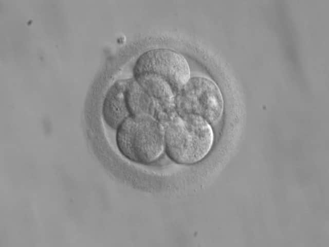
Beyond the number of cells, embryologists look for additional signs of a healthy embryo, such as: fragments, evenly sized cells, and not multinucleated. This is how an embryologist “grades” an embryo. Most of the time, fertility clinics don’t grade embryos until they are around 48 hours old. Grading embryos helps determine which embryos have the best chance of implantation and a healthy, live birth after IVF pregnancy.
Embryo Transfer Process Overview
| Step 1. Scheduling |
| The fertility clinic schedules embryo transfer at the time the fertilized eggs become ready to transfer. |
| Step 2. Preparation |
| The fertility clinic and staff prepare the embryos to be transferred. Typically, a fertility doctor places embryos in a syringe for use during transfer. Before this happens, the clinic and the couple or woman will discuss how many embryos to transfer and what to expect. |
| Step 3. Sedation |
| Generally, many women do not need anesthesia or sedation during embryo transfer. The process should be painless. However, some women may require sedation.The fertility clinic may use a light sedative for comfort. |
| Step 4. Transfer |
| The fertility doctor transfer the embryos to the uterus using the catheter. |
| Step 5. Rest |
| Sometimes, women will rest for a period of time before leaving the clinic. However, rest isn’t required for successful transfer. The clinic typically sends couples and women home with follow up instructions for what to expect after the transfer. |
When is an embryo transfer procedure scheduled?
As long as the embryos develop normally, embryo transfer takes place 2 to 4 days after egg retrieval and fertilization. Some fertility clinics and their patients choose to wait longer than that, when an embryo becomes a blastocyst.
On the other hand, if the fertility clinic is using frozen embryo transfer (FET), the timing of the transfer will depend largely on the patient’s cycle and schedule with the fertility clinic.
During this time, the fertility clinic and patient keep in close communication with one another. If embryos do not develop (i.e. if eggs do not become fertilized), the clinic will discuss further options with the patient. Similarly, the clinic may perform additional procedures before transfer, such as assisted hatching, to help the embryo implant.
Also, prior to transfer, if the patients have opted to do any genetic screening, the embryologists will perform the tests on the embryos.
What happens during an embryo transfer procedure?
Embryos must be transferred with extreme care and consideration. At every step of the process, embryologists and fertility doctors must handle the embryos properly; otherwise, the risk of a failed transfer increases. The uterus and the transfer itself should be monitored by ultrasound. And, most importantly, the fertility doctor has to take great care not to damage the lining of the uterus or the embryos during transfer.
The following steps break down the end to end embryo transfer procedure. While every fertility clinic is different and may have a unique process, the below steps provide a high-level overview of the process that most clinics use. The differences in procedure mainly have to do with patient and embryo identification procedures and check-in processes.
- After an embryologist determines that embryos are ready for transfer, the fertility clinic contacts the patient to arrange a time for transfer.
- Upon arrival, patients check-in and follow standard clinic processes for identification. Clinics must be extremely careful that embryos and patients match, and that no misidentification happens.
- Patients and their doctors discuss how many embryos will be transferred and how many will be frozen (if applicable). At this point in time, patients complete documentation regarding the procedure and number of embryos to transfer.
- Fertility specialists load the embryos to be transferred into a syringe.
- If the patient receives any sedatives, they’re administered at this time.
- The doctor carefully inserts a catheter through the vagina into the uterus. A catheter is a small, hollow, plastic tube.
- The embryos are loaded into the catheter and then pushed into the uterus. Ultrasound technology helps doctors and embryologists ensure that embryos were placed inside the uterus. The placement of the embryos also matters a great deal.
- The doctor removes the catheter slowly to avoid causing uterine contractions or damaging the lining in any way.
- From here, the patient usually rests for a small period of time before the fertility doctor dicscharges her.
Usually, patients do not need recovery time from the embryo transfer – especially if the embryos were frozen prior to transfer. However, it’s possible that recovery time from the egg retrieval process overlaps a fresh embryo transfer. So, some patients will still need to recover as it can take up to two weeks for complete recovery from egg retrieval.
Are there any risks with an embryo transfer?
Even though the transfer itself is very brief, there are some risks. Choose a clinic with a strong reputation for successful transfers, because it’s the most critical procedure in the IVF treatment.
Some risk is simply inherent in the process, due to how fragile embryos are. Fertility clinic staff can unknowingly damage embryos at any point in time. Additionally, embryos don’t survive very long outside of the womb. So clinicians and embryologists have to work quickly and carefully.
A small amount of risk stems from the placement of the embryos in the uterus, also. Research shows that embryos placed in the middle of the uterus have the best chance at implantation. So, doctors should carefully monitor the placement of the catheter and location of the transfer using high quality ultrasound machines.
The highest risk is the potential for a failed transfer. Failed embryo transfer means the embryos did not implant – either because they did not survive long enough, did not divide, or for another reason. Embryo transfers fail for a number of reasons; however, the primary causes of a failed transfer involve the lining of the uterus.
Most of the time, fertility specialists won’t be able to pinpoint the exact reason for failure. Possibly, a number of things caused embryo transfer failure. Very good clinics take precautions to try and limit the potential for failure. To reduce risk, fertility doctors give medications to thicken the uterine lining, and of course, carefully handle the embryos during transfer.
After the Embryo Transfer
Once the clinic transfers the embryo successfully, IVF treatment is technically over, and at this point in time, everyone – the patient and the fertility specialist – must allow nature to take over.
The transfer is the last step in the IVF treatment process. However, for patients, the embryo transfer is really just the beginning! After transfer, patients return home with any instructions from the clinic for rest and follow up. The clinic typically schedules a pregnancy test (blood test) for fourteen days after transfer. They use the blood test to determine pregnancy.
But before that, 4 to 5 days after transfer, in a normal pregnancy, the embryos should successfully implant in the lining of the uterus. There, they begin to divide even further and continue their development from an embryo to a fetus.
If a woman is pregnant, she may notice light or dark brown spots, or implantation spotting, during this time. That is very normal; however, patients should always contact the fertility clinic immediately should any questions or concerns come up.
Frequently Asked Questions
Is embryo transfer painful?
No. The patient may experience some very mild discomfort while the doctor inserts the catheter into the vagina. However, most of the time, doctors do not use any anesthesia at all and may only use a light sedative.
How long does embryo transfer take?
Technically, embryo transfer takes only 5 to 10 minutes. However, patients should expect to be at the clinic for much longer than that. A typical visit includes patient and embryo identification/matching, a discussion about the embryos and how many to transfer, the transfer procedure itself, and a short period of rest.
References
- Embryo Transfer – American Pregnancy Association (http://americanpregnancy.org/infertility/embryo-transfer/)
- In Vitro Fertilization (IVF) What You Can Expect – Mayo Clinic (http://www.mayoclinic.org/tests-procedures/in-vitro-fertilization/details/what-you-can-expect/rec-20206943)